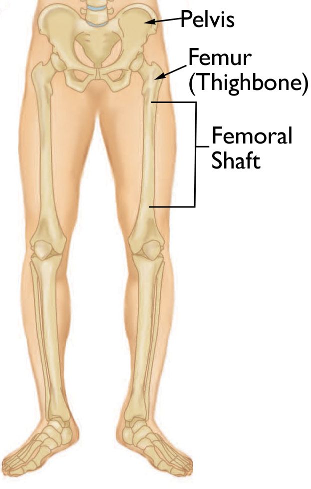
The femur bone is the body’s strongest and longest bone. It’s situated between the hip and knee joints in the lower limb. The femur’s shape is unique. It’s ideal for supporting the various muscular and ligamentous connections in this area. It also extends the limb to its maximum potential during ambulation. The femur connects to the pelvic bone proximally. It collaborates with the patella and the proximal prospect of the tibia from a distance.
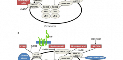
The route of endochondral ossification begins to evolve the femur during the 5th and 6th gestational week. There bones are composed using a cartilage-dependent foundation. Thus many ossification sites (bone growth points) occur during pregnancy. But the bone continues to develop throughout infancy and early manhood. Between the ages of 14 and 18, the ossification of the femur is completed.

Femur Bone Definition
The femur is also known as the thighbone. It is the upper or hind leg’s bone. The head and hip (at the acetabulum) create a ball-and-socket joint. It completes the head kept in place by a ligament (ligamentum teres femoris). Femur stays within the socket with strong surrounding ligaments. The femur’s neck links the shaft and head at a 125° angle in humans. This is ideal for walking. The gluteus medius and minimus muscles connect to a protrusion of the femur on the outer top of the thigh. The linea aspera, a bone pillar, strengthens the back of the shaft. This slightly convexes forward.
In humans, the femur is long and thin or fragile. In great apes, the femur is shorter, more curved, and more robust. The absence of a ligamentum teres femoris in orangutans allows for virtually full rotational motion of the lower limb. But this comes at the expense of strength and stability.
Femur Bone Location in Body
The femur is a bone that is located in the thigh. It is the body’s biggest bone and the only one in the upper leg. The femur is a long bone in the human body. Long bones, short bones, flat bones, and irregular bones are the four kinds of bones. Long bones have spongy bones at both ends. They have hollows filled with bone marrow in the shaft. They are longer than they are broad. A mature male’s femur is around 19 inches long. And it’s a little less than an inch in diameter. The head of the femur is the proximal end of the femur (the portion nearest to the heart).
Read Also: Capacitance: Units & Formula
The neck and the greater trochanter are located below the head of the femur. However, the tendons that link the gluteus minimus and gluteus medius muscles are attached to the greater trochanter. These muscles also aid in walking and running by pulling the leg. An extension of the leg or hip is also what this is called.
The lesser trochanter is located at the base of the femur’s neck, below the larger trochanter. The lesser trochanter is a portion of the femur. It’s in connection with a pair of muscles that aid in thigh flexion (lift the leg forward). The gluteal tuberosity is located below the lesser trochanter. And it is where the gluteus maximus attaches.

Femur Bone Anatomy
Proximal Femur
The femoral neck connects the bulbous femoral head to the shaft of the femur at the proximal end of the femur. The apex of the major trochanter is also at the same level as the femoral head’s centre. Under the femoral neck is the lesser trochanter. It runs along the femoral body’s inner rear surface. At this moment, the iliopsoas muscles might also connect.
The femoral head’s centre and the acetabulum’s centre are in perfect alignment. At the apex of the femoral head, the fossa capitis femoris is in position medially. The ligamentum teres is connected there. At the point where the body and the femoral neck meet, the greater trochanter intersects. The abductor muscles are also attached to it.
The Inclination Angle
From the front, the angle between the femoral neck and the medial side of the femoral shaft. Due to loading pressures over the femoral neck in weight-bearing and walking. It is greater at birth but diminishes during childhood and teenage growth. A normal angle of inclination of 125 degrees enhances femur mobility at the hip joint. It does so by putting the femoral head and neck in a biomechanically favourable position for walking. Coxa vara less than 125 degrees; coxa valga higher than 125 degrees are abnormal angles. Both change the biomechanics of the hip, resulting in misalignment and problems.
Shaft
The body of the femur is the primary shaft of the femur. The patella (knee cap) and the lower leg bones, the tibia and fibula, are all connected. They get together at the distal end of the femur. A saddle on the distal end of the femur sits on the top of the tibia. The condyles are the rounded margins on either side of the knee joint. The patellar groove is the dip between the condyles.
Linea Aspera (roughened bone ridges) stays on the rear surface shaft. The medial and lateral supracondylar lines arise when they divide inferiorly. Between them is the smooth popliteal surface. The pectineal line runs parallel to the medial edge of the linea aspera. The gluteal tuberosity, which is where the gluteus maximus attaches, is formed by the lateral border.
The linea aspera expands and creates the popliteal fossa’s floor, while the medial and lateral borders form the medial and lateral supracondylar lines. At the adductor tubercle, where the adductor Magnus connects, the medial supracondylar line terminates.
Medullary Body
The medullary cavity contains bone marrow. It is located within the femur’s body. There are solid regions of compact bone at the extremities of the femur. However, these do not contain marrow. Spongy bones also surround the compact bone. Compact Bones also have many tiny cavities distributed throughout them.
Muscles surrounding Femur Bone
The anterior, medial, and posterior compartments of the thigh muscles are split. The gluteal compartment can also split. The anterior compartment houses the femur.
- The muscles of the anterior compartment are mostly employing themselves. Thus they are working for hip flexion and knee extension. The pectineus, iliopsoas, and sartorius muscles are also hip flexors.
- Leg adduction is the primary function of the medial compartment. The adductor long, adductor brevis, adductor Magnus, gracilis, and obturator externus are also together. They are all part of this muscle group.
- Hip extensors and knee flexors make up the majority of the muscles. The bicep femoris, semitendinous, and semimembranous muscles make up this muscle group.
- Segment of the gluteus maximus. The gluteus maximus, medius, and minimus make up the superficial layer. The major functions of the superficial gluteal include hip extension, abduction, and internal rotation. The piriformis, obturator internus, quadratus femoris, and superior and inferior gemellus make up the deep layer.
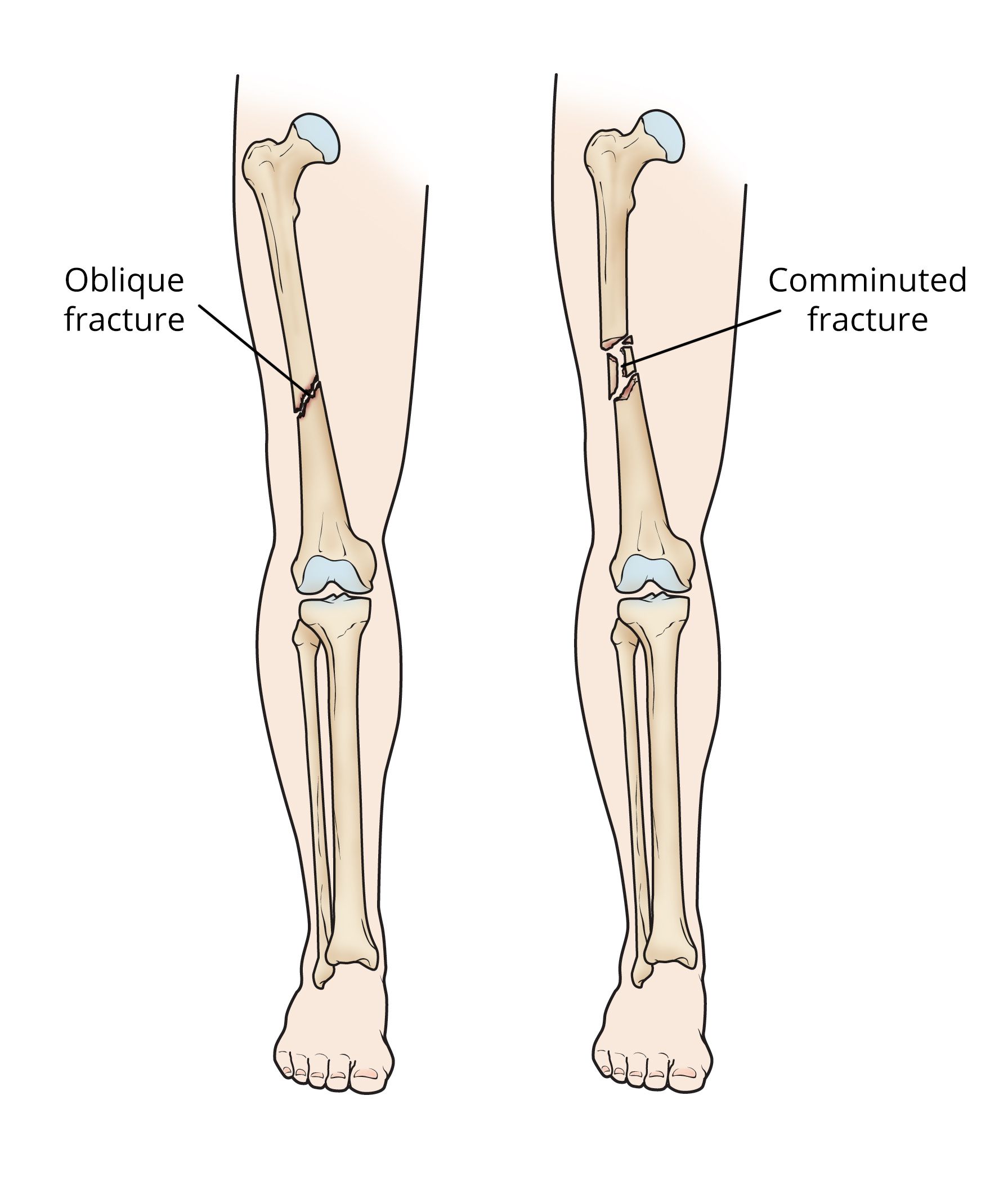
Femur Bone Fractures and Injuries
Fractures are the most frequent femur ailment. Though a femur may break with a lot of force. However, specific parts of the femur are more vulnerable. The most susceptible part of the femur is the neck. Fractures in the elderly are more likely as bone density declines with age.
NOFX (neck of femur fractures) are becoming more prevalent in the older population. This is due to low-energy falls in the presence of osteoporotic bone. Women are more likely to have them. They commonly develop in younger individuals as a result of high-energy incidents.
Perthes disease
Perthes disease is an uncommon hip joint condition that affects children. Blood flow to the head of the femur is easily disturbable. When the head of the femur stops moving, the bone tissue dies due to a lack of blood supply. This condition is known as osteonecrosis.
Patellofemoral pain syndrome
Pain around or behind the patella, usually with an insidious start, is a frequent sports-related ailment. The aetiology of the disease is unknown, although it might be due to neurologic, genetic, neuromuscular, and biomechanical causes.
Diagnosis of a broken femur
The doctor will begin by taking an X-ray. They may also request a CT scan if they ask for further information. A fractured femur is uncommon since the femur is such a tough bone. The healing process can also take up to six months. It is also devidable into four stages:
- The body begins to repair itself.
- Inflammation occurs throughout the body.
- With new bone development, the body regenerates.
- Mature bone is also replacable by freshly produced bone when the body remodels.
Surgery and medicines are important for the majority of fractured femurs.
Various procedures, both internal and external, are there to keep the bones in place while they recover. Intramedullary nailing is the most frequent procedure for a fractured femur. However, this procedure involves inserting a rod into the length of the bone. It needs to get security in place using screws above and below.
Following a femur break, the doctor will most likely suggest physical therapy. It helps to strengthen the muscles around the bone. Once the bone puts itself back into its normal place, it gets stable. Exercises to strengthen the thigh will also aid in the leg’s restoration.